Home

Cottage Miner Mediterranean Sea break apart fish probes cooperate Search Exclusion

Figure 2 from FISH analysis for the detection of lymphoma-associated  chromosomal abnormalities in routine paraffin-embedded tissue. | Semantic  Scholar
Figure 2 from FISH analysis for the detection of lymphoma-associated chromosomal abnormalities in routine paraffin-embedded tissue. | Semantic Scholar

MYC Break Apart FISH Probe Kit - CytoTest

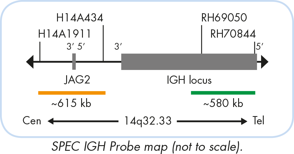
ZytoLight SPEC IGH Dual Color Break Apart Probe - ZytoVision GmbH

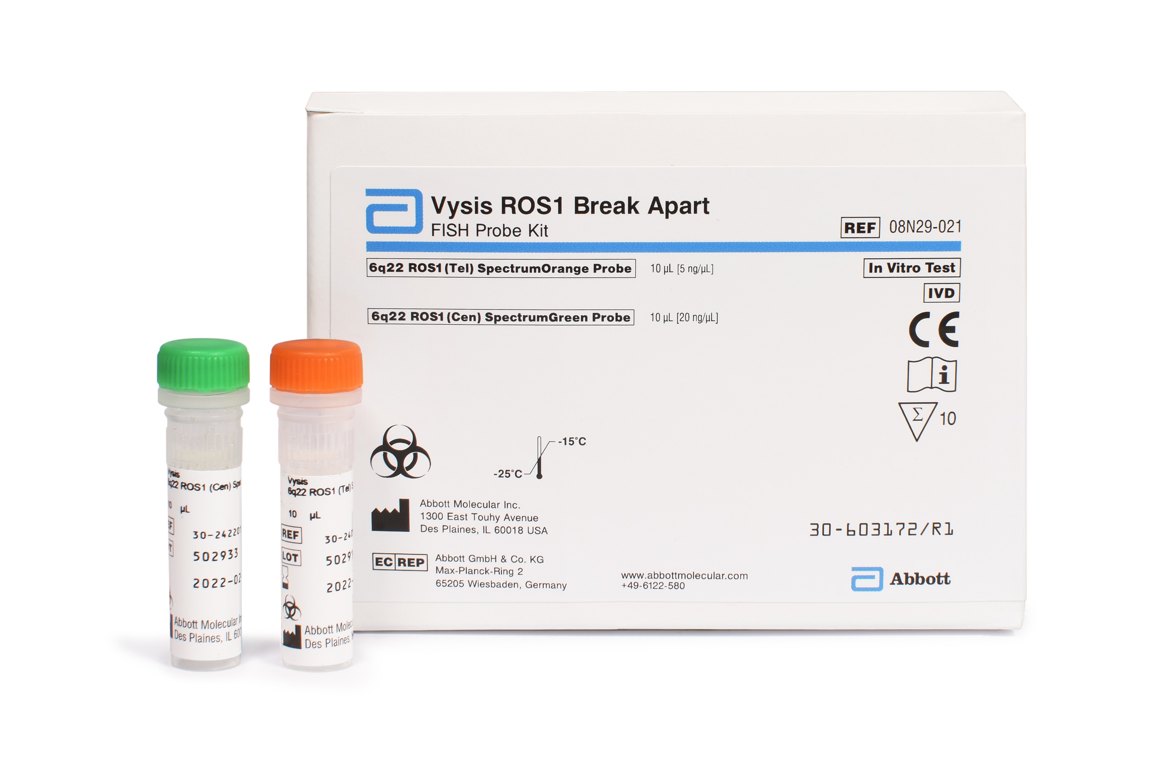
Vysis ROS1 Break Apart FISH Probe Kit | Abbott

FISH analysis for the detection of lymphoma-associated chromosomal  abnormalities in routine paraffin-embedded tissue. - Abstract - Europe PMC
FISH analysis for the detection of lymphoma-associated chromosomal abnormalities in routine paraffin-embedded tissue. - Abstract - Europe PMC

Schematic representation of the dual-color probe FISH break-apart assay...  | Download Scientific Diagram
Schematic representation of the dual-color probe FISH break-apart assay... | Download Scientific Diagram

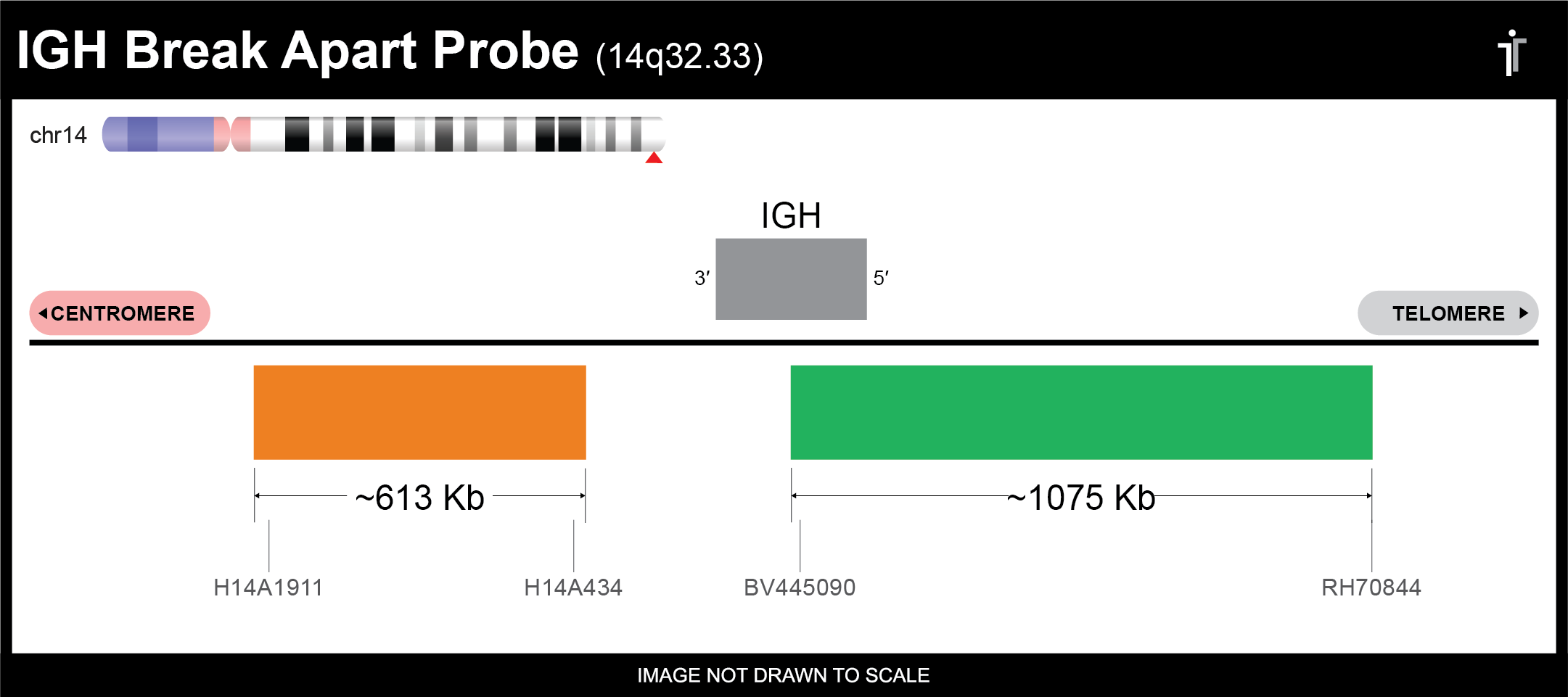
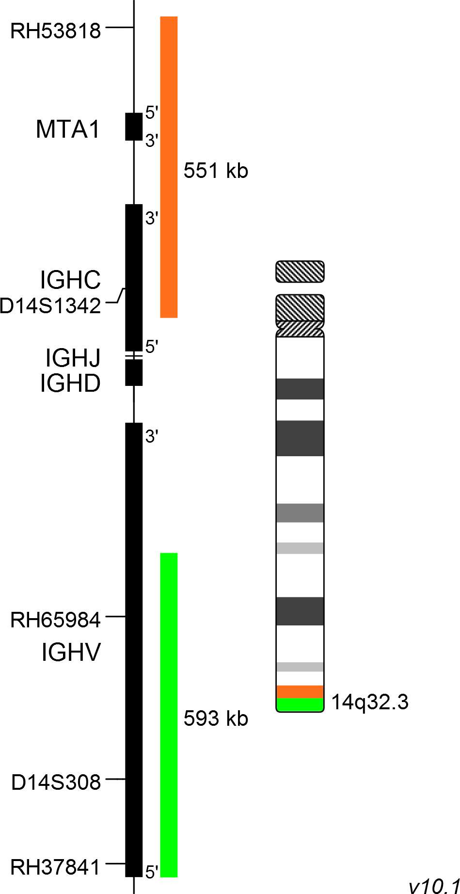
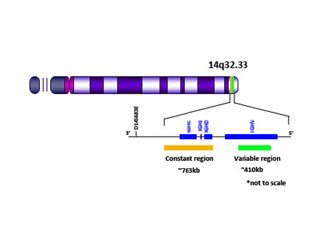
IGH Break Apart FISH Probe

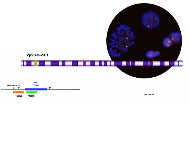
Test the new ALK probe for FISH automation Clinisciences

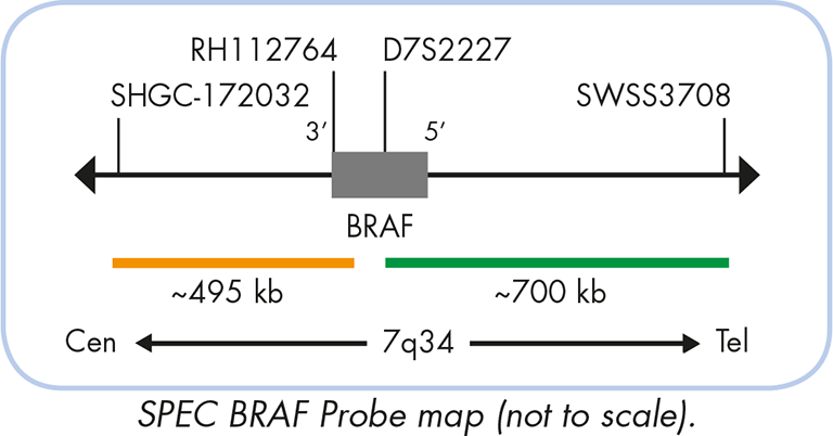
ZytoLight SPEC BRAF Dual Color Break Apart Probe - ZytoVision GmbH

Vysis LSI MALT1 Break Apart FISH Probe Kit

New Reflex Fish Procedure For Large Cell Lymphoma and Change To BCL2 Break  Apart Probe - YouTube
New Reflex Fish Procedure For Large Cell Lymphoma and Change To BCL2 Break Apart Probe - YouTube

MYC break-apart FISH probe set reveals frequent unbalanced patterns of  uncertain significance when evaluating aggressive B-cell lymphoma | Blood  Cancer Journal
MYC break-apart FISH probe set reveals frequent unbalanced patterns of uncertain significance when evaluating aggressive B-cell lymphoma | Blood Cancer Journal

ALK Break Apart FISH Probe Kit - CytoTest

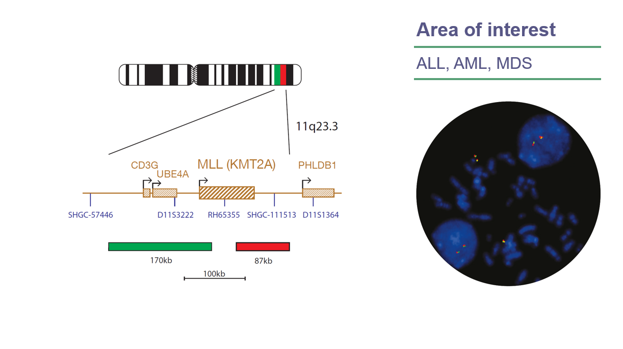
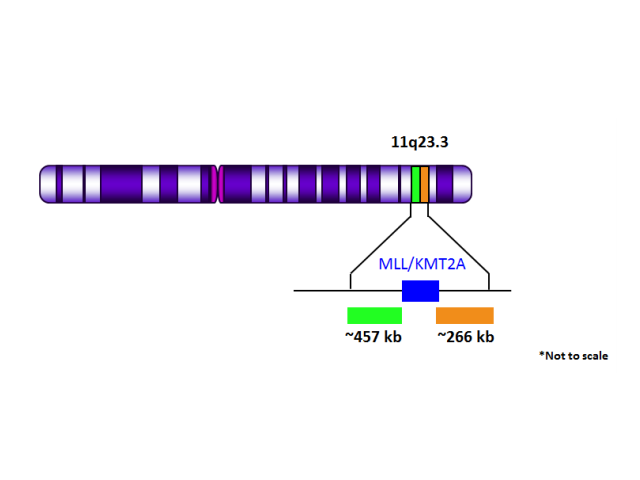
CytoCell MLL (KMT2A) Breakapart FISH Probe | OGT

MLL Break Apart (Orange/Green) FISH Probe - Biocare Medical

RET IQFISH Break-Apart Probe (Dako Omnis) | Agilent

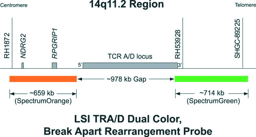
Vysis TRA/D Break Apart FISH Probe Kit

Common FISH signal patterns | OGT

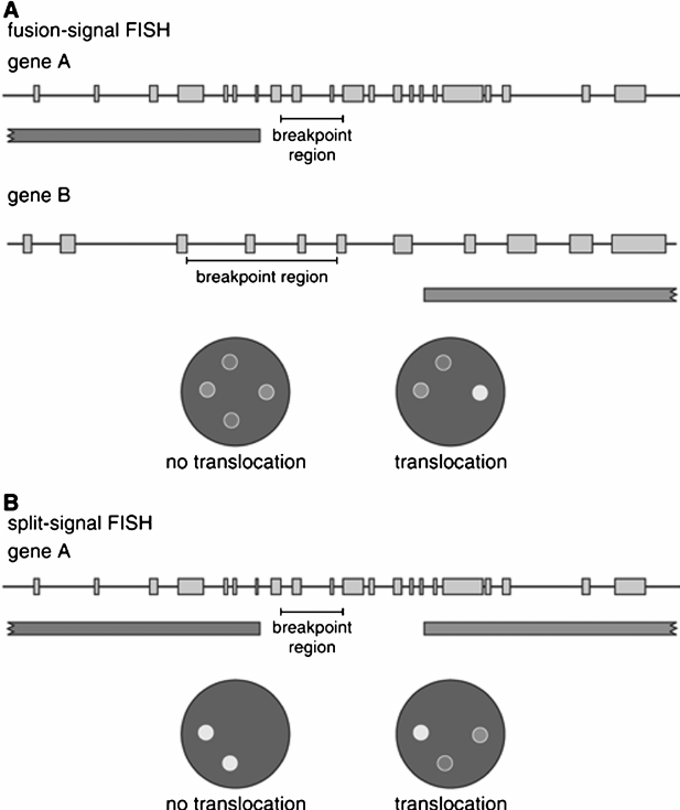
Differences between fusion-signal FISH and split-signal FISH. A.... |  Download Scientific Diagram
Differences between fusion-signal FISH and split-signal FISH. A.... | Download Scientific Diagram

XL IGH BA - Break Apart Probe | MetaSystems Probes

TFE3 Break Apart FISH Probe

ALK (2p23.2) Break Apart FISH Probe Orange/Green - Biocare Medical

Schematic design of the break-apart FISH assays and exemplary findings....  | Download Scientific Diagram
Schematic design of the break-apart FISH assays and exemplary findings.... | Download Scientific Diagram

BRAFBA-20-REGO | BRAF Break Apart FISH Probe, Dye Color: Red-Gold

IGH (14q32) Break Apart FISH Probe Orange/Green - Biocare Medical

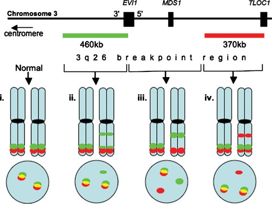
A simple FISH assay for the detection of 3q26 rearrangements in myeloid  malignancy | Leukemia
A simple FISH assay for the detection of 3q26 rearrangements in myeloid malignancy | Leukemia

8. FISH with break-apart probes to determine the presence of a... |  Download Scientific Diagram
8. FISH with break-apart probes to determine the presence of a... | Download Scientific Diagram
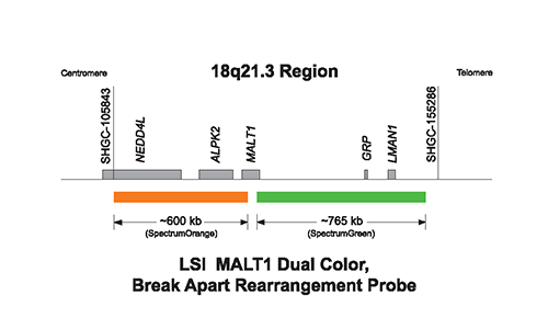
Figure 1 from FISH analysis for the detection of lymphoma-associated  chromosomal abnormalities in routine paraffin-embedded tissue. | Semantic  Scholar
Figure 1 from FISH analysis for the detection of lymphoma-associated chromosomal abnormalities in routine paraffin-embedded tissue. | Semantic Scholar